Session 3A: 10:00 AM – 12:10 PM Pacific Time on Friday, August 21.
Jong Seto*, Behzad Rad, Paul Ashby
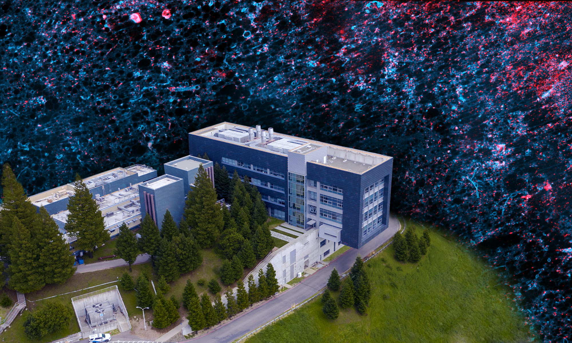
The cell is a dynamic environment with chemical and mechanical processes happening at the nanoscale and over a broad range of timescales. Recent advances in characterization techniques have enabled detailed in situ investigations to probe these processes. Methods including super resolution microscopy, light-sheet microscopy, scanning probe microscopy, cryo-electron microscopy and synchrotron X-ray techniques can capture processes in real time and under physiologic conditions. In situ characterization tools will play an increasingly essential role in gaining fundamental understanding of cellular processes, and multiple complementary, correlative techniques will be required. This symposium will bring together researchers involved in cross-disciplinary work taking place at the Molecular Foundry and beyond to showcase cutting edge multimodal in situ capabilities and foster collaboration and new directions.
Symposium Schedule:
10:00 am
Invited: Timing the process of bacterial division with nm spatial and ms temporal precision
Prof. Georg Fantner, EPFL
10:30 am
In situ Imaging of Hyphal Growth in Candida albicans via Atomic Force Microscopy
Prof. Mehmet Baykara, Mechanical Engineering, UC Merced
10:40 am
Structural and mechanical characterization of tissue-like bacterial assemblies using In-situ atomic force microscope
Dr. Dong Li, Molecular Foundry, Berkeley Lab
10:50 am
10-minute break
11:00 am
Invited: Single-molecule imaging of cells detecting nutrients in their local environment
Prof. Julie Biteen, Departments of Chemistry and of Biophysics, University of Michigan
11:30 am
Multifunctional neural probes for simultaneous readout and on-demand passive stimulation of in vivo neural networks
Dr. Vittorino Lanzio, Molecular Foundry, Berkeley Lab
11:40 am
Invited: High speed, high resolution 3D imaging of biomaterials with synchrotron X-ray microCT
Dr. Dula Parkinson, Advanced Light Source, Berkeley Lab
Symposium Abstracts:
10:00 AM
Timing the process of bacterial division with nm spatial and ms temporal precision structure and dynamics
Prof. Georg Fantner
EPFL
Several processes have to occur in a cell before it can divide, many of which are still only partially understood. Processes such as FtsZ ring formation, chromosome separation, membrane synthesis and cytokinesis have each been studied individually in great detail, often using static methods. Establishing the sequence and possible dependence of these processes however requires time resolved live-cell imaging, at high resolution. Here we present a concise time-sequence of events in the division of Mycobacterium Smegmatis, obtained using a combination of multi-day time-lapse atomic force microscopy (AFM) and time-lapse fluorescence microscopy. By combining the nanoscale 3D information from AFM and the biochemical specificity from fluorescence microscopy we demonstrate that the location of future cell division in M. Smegmatis is determined up to several generations before cell birth, and that geometric factors may guide the localization of FtsZ rings. We characterized the cell division from the early stage pre-selection of division cites, to assembly and subsequent disassembly of the FtsZ ring, localization of Wag31 and cytokinesis all the way to the rapid cell separation, and subsequent onset of cell pole growth. The nanometer spatial resolution of time-lapse AFM imaging of living bacteria shows us that even the most fundamental processes in “simple” organisms still hold many surprises.
10:30 AM
In situ Imaging of Hyphal Growth in Candida albicans via Atomic Force Microscopy
Prof. Mehmet Baykara
Mechanical Engineering, UC Merced
Coauthors: Arzu Çolak, Mélanie A. C. Ikeh, Clarissa J. Nobile
Candida albicans is an opportunistic fungal pathogen of humans known for its ability to cause a wide range of infections. One major virulence factor of this fungus is its ability to form hyphae that can invade host tissues and cause disseminated infections. Here we introduce an atomic-force-microscopy-based method to investigate C. albicans hyphal growth in situ on silicone elastomer substrates, a clinically-relevant material. It is shown that hyphal growth rates differ significantly for measurements performed at different temperatures. Furthermore, the growth of hyphal cells is explored under the influence of caspofungin and fluconazole, the most widely prescribed antifungal drugs. It is found that fluconazole is more effective than caspofungin in suppressing hyphal growth rates. Finally, the effects of fluconazole and caspofungin on the mechanical properties of hyphal cell walls are investigated using force-distance spectroscopy. An increase in Young’s modulus and a decrease in adhesion force are observed in hyphal cell walls subjected to caspofungin treatment. Young’s moduli are not significantly affected in hyphal cell walls following a treatment with fluconazole; the adhesion force, however, increases.
10:40 AM
Structural and mechanical characterization of tissue-like bacterial assemblies using In-situ atomic force microscope
Dr. Dong Li
Molecular Foundry, Berkeley Lab
Coauthors: Victor Mann1, Marimikel Charrier1, Paul Ashby1, Kathleen Ryan2, Caroline Ajo-Franklin3, Behzad Rad1
1Berkeley Lab; 2UC Berkeley; 3Rice University
Engineered Living Materials (ELMs) that incorporate genetically modified cells to actively adjust the expression and organization of biomacromolecules are excellent candidates for applications in bioelectronics, biocatalysis, and smart materials. However, it is challenging to perform in-situ characterization on ELMs with high temporal and spatial resolution, due to their dynamic chemical, and mechanical properties. In this talk, I will present our new strategy to create living bacterial tissues using a genetically modified bacterium: Caulobacter crescentus, and how we use in-situ atomic force microscope (AFM) to obtain both the surface morphology and viscoelasticity of this new ELM. The surface layer (S-layer) protein of C. crescentus has been engineered to display a 2D array of functional peptide, i.e. SpyTag, over the entire cell body. A layer of tightly packed nanoparticles is attached to the recombinant protein lattice through formation of SpyCatcher-SpyTag iso-peptide bond. The functionalized bacterial cells are further crosslinked into hierarchically ordered, tissue-like assemblies. The bacterial tissues have high stiffness and can self-regenerate after being damaged due to the continuous growth of C. crescentus cells and the expression of new S-layer proteins.
11:00 AM
Single-molecule imaging of cells detecting nutrients in their local environment
Prof. Julie Biteen
Departments of Chemistry and of Biophysics, University of Michigan
Single-molecule microscopy accesses nanometer-scale information on a benchtop microscope, providing a platform to super-resolve real-time, subcellular dynamics in living cells. We have been developing new single-particle tracking methods to locate, track, and analyze single-molecule data to answer fundamental, unanswered questions in microbiology. I will discuss how these direct, quantitative, and high-resolution approaches are enabling us to measure and understand the dynamical interactions essential for catabolizing carbohydrates and adapting to a changing landscape of nutrients in the human gut microbiome. Overall, our results achieve fundamental insight of relevance to human health and disease, and our work offers algorithms that can be further applied to other problems in chemistry, biology, and biophysics.
11:30 AM
Multifunctional neural probes for simultaneous readout and on-demand passive stimulation of in vivo neural networks
Dr. Vittorino Lanzio
Molecular Foundry, Berkeley Lab
Coauthors: Gregory Telian2, Alexander Koshelev3, Paolo Micheletti1, Gianni Presti1,4, Elisa D’Arpa1,5, Paolo De Martino1,5, Monica Lorenzon1, Peter Denes1, Melanie West1,2, Simone Sassolini1, Scott Dhuey1, Hillel Adesnik2, Stefano Cabrini1
1Berkeley Lab; 2UC Berkeley; 3Abeam Technologies, Inc.; 4Politecnico di Milano; 5Politecnico di Torino
Neural probes are invasive brain devices for the in vivo interrogation of neural cells in networks with high spatial and temporal resolution (single neuron and single neural events, respectively).
At the Molecular Foundry, we fabricate these devices and integrate multimodal micro/nanocircuits to simultaneously read out and manipulate neural activity. We achieve a readout of a large number of neurons (~50) through arrays of sensors and a simultaneous manipulation by integrated photonics, which shine light to activate neurons. Specifically, the integrated photonics deliver light in the area of interest – to activate the desired groups of neurons along the length of the device – using ring resonators, which act as passive optical switches.
Thus, with these devices, we aim at analyzing the contribution of groups of neurons in networks by measuring their activity and by (simultaneously) selectively activating them. In this talk, I will describe these devices, their design, simulation, and preliminary in vivo validation towards the advancement of the neuroscientific field and our understanding of neural network functioning.
11:40 AM
High speed, high resolution 3D imaging of biomaterials with synchrotron X-ray microCT
Dr. Dula Parkinson
Advanced Light Source, Berkeley Lab
Coauthors: Craig Brodersen1, Claire Acevedo2, Andrew McElrone3
1Yale University; 2University of Utah; 3UC Davis
The high flux of synchrotron beamlines allows high speed, high resolution 3D imaging. The penetrating power of X-rays allows the study of internal structures in situ and even inside of sample environments with minimal sample preparation. I will describe a selection of projects that have used the hard X-ray microtomography beamline of the Advanced Light Source, in combination with other methods, to reveal new details of biological processes. First, I will describe studies of cellular organization in stems and leaves of living plants, and interplay with water and gas transport. Second, I will describe work on bone structure and fracture mechanics, and how they are influenced by different factors. Finally, I will describe our efforts to use machine learning and high performance computing to extract understanding from the petabytes of phenotype data.
